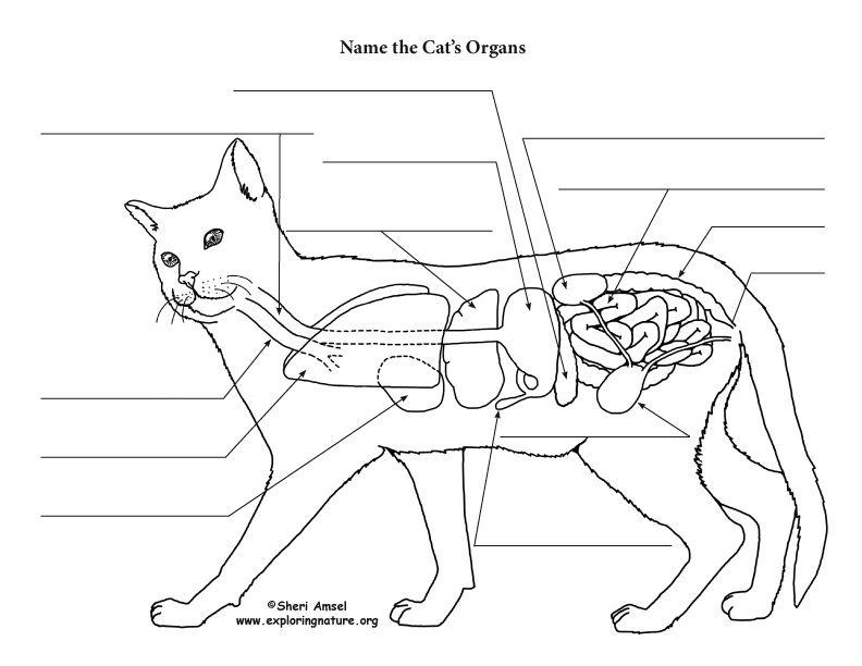
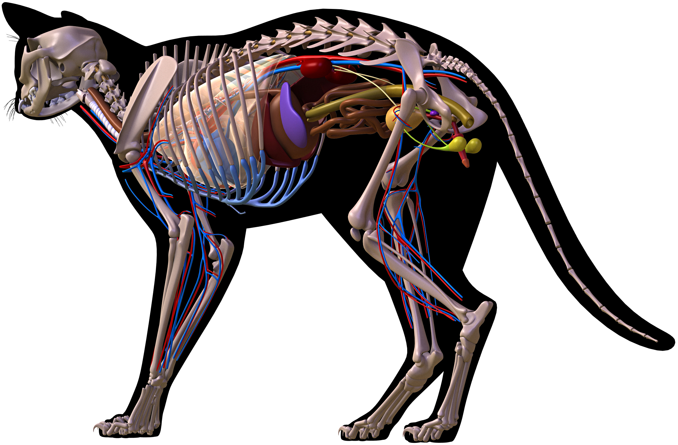
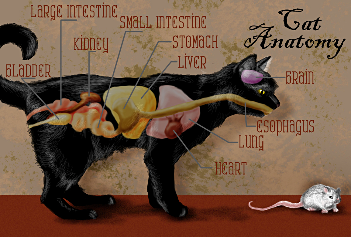
Cat Anatomy (Thoracic and Abdominal Organs)
Computed tomography ( CT or CAT scan) is one of the most commonly used medical imaging procedures in clinical practice, along with radiography (x-ray) and magnetic resonance imaging (MRI).
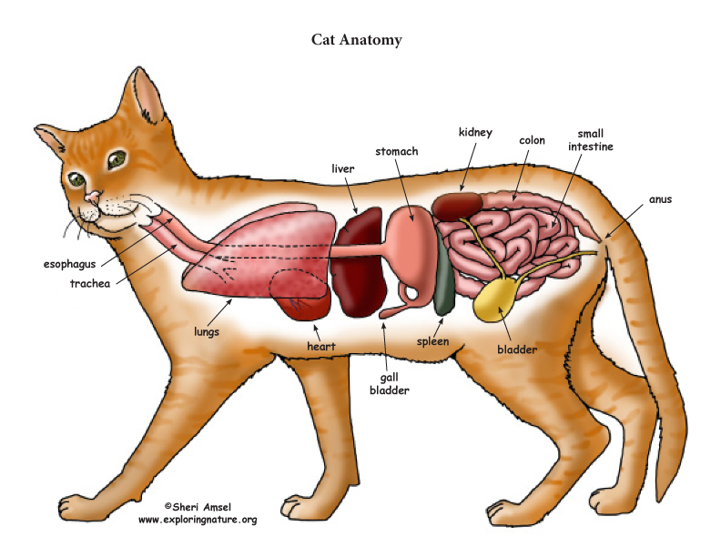
Cat Anatomy (Thoracic and Abdominal Organs)
The aim of the present review is to describe the anatomy of the gastrointestinal tract of the abdomen in cats in combination with the surgical procedures that are performed in each region, highlighting the points of surgical interest.

5.3 Cat Musculature Medicine LibreTexts
Cat anatomy comprises the anatomical studies of the visible parts of the body of a domestic cat, which are similar to those of other members of the genus Felis . Mouth Sharp spines or papillae found in a cat's tongue. 5 types of papillae can be found in the dorsal aspect of the tongue: filiform, fungiform, foliate, vallate, and conical.
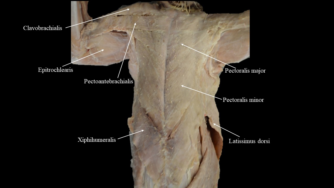
5.3 Cat Musculature Medicine LibreTexts
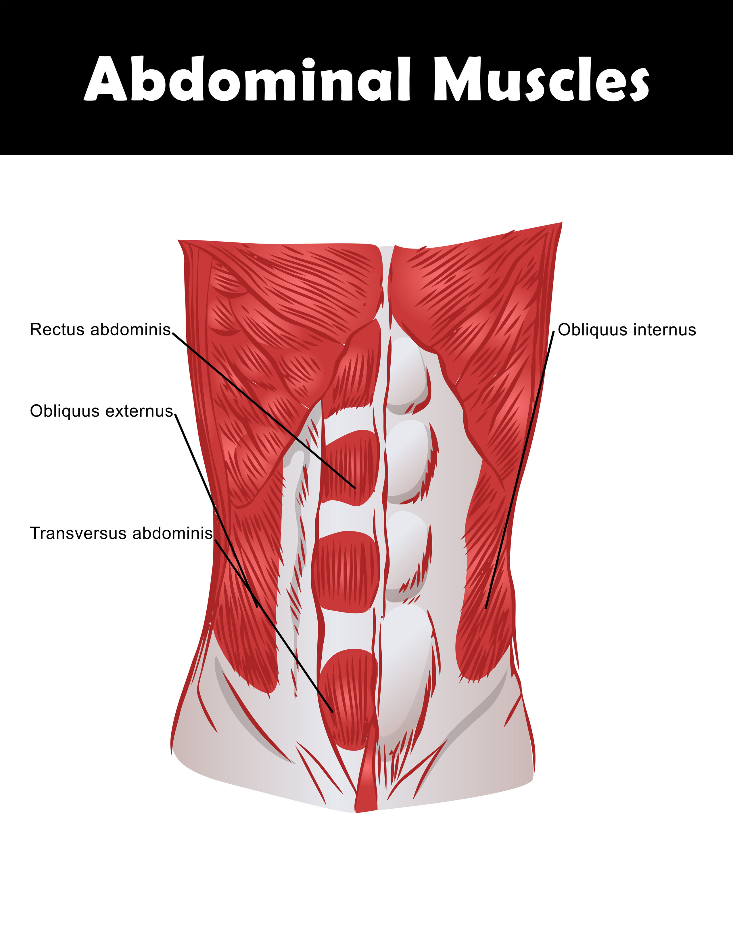
The cat muscle anatomy includes the origin, insertion, and fiber direction of every single muscle from the different regions of the body. Here, I will show you the essential muscles from the face, neck, forelimb, abdomen, and hindlimb. You will also find the description of these muscles from the different regions.
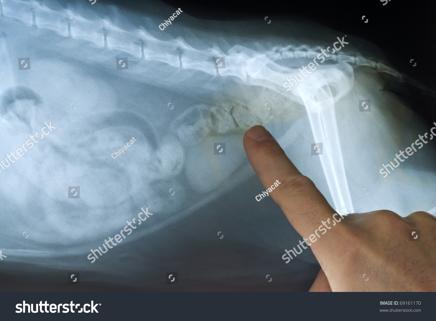
Cat Abdominal XRay Stock Photo 69161170 Shutterstock
June 16, 2021 | Issue: July/August 2021 Krysta Janas DVM Karen Tobias DVM, MS, DACVS Yakov Oskanov/shutterstock.com Exploratory laparotomy or celiotomy is commonly performed for diagnosis, treatment, or prognostication of traumatic, inflammatory, infectious, neoplastic, and congenital abdominal conditions.

Muscles of the Abdomen and Ribs Laminated Anatomy Chart Anatomie
Coronal Bone EXAMPLE REPORTING TEMPLATE WITH CHECKLIST: LOWER CHEST: Lung bases are clear. No pleural or pericardial effusion Lung bases Pleural effusion Pericardial effusion LIVER AND BILIARY: Normal liver morphology and enhancement. No masses. Normal gallbladder morphology. Normal caliber intrahepatic and common bile ducts. Morphology Enhancement

anatomy of the abdominals
Radiology basics of abdominal CT anatomy with annotated coronal images and scrollable axial images to help medical students and junior doctors learning anatomy.. Axial CT abdomen (to scroll - click and drag the image up or down) Transabdominal ultrasound views Longitudinal right flank. Ultrasound probe position.
Links to Pictures on the Physiology of Cats
Atlas of CT Anatomy of the Abdomen This photo gallery presents the anatomy of the abdomen by means of CT (axial, coronal, and sagittal reconstructions). Click a link to get Axial view - Coronal view - Sagittal view < > Abdominal Computed Tomography

Abdominal ultrasound anatomy Small Animal Ultrasonography
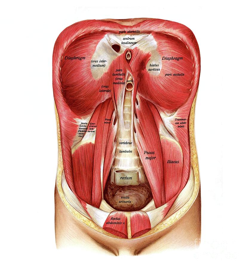
right colic vessels. right common iliac artery. abdominal portion of the ureter. left colic artery. umbilicus. ileocecal valve. left common iliac artery. quadratus lumborum muscle. transversus abdominis muscle.

Anatomía del gato, Anatomía del perro, Anatomia veterinaria
Examination Modern veterinary medicine has a much better understanding of a cat's digestive system and your trusted DVM veterinarian will carry out a full assessment of your troubled cat. The science of veterinary medicine now has a detailed understanding of the workings of the digestive tract.

Cat musculature Atlas of Comparative Vertebrate Anatomy
25/04/2023 28/05/2022 by Sonnet Poddar The cat digestive system includes a mouth cavity, pharynx, alimentary canal, and different accessory organs. There are two major divisions in the mouth cavity of a cat - vestibule and mouth cavity proper. The alimentary canal of a cat starts with the esophagus and ends at the large intestine.
abdomen internal structure
We created an anatomical atlas of abdominal and pelvic CT which is an interactive tool for studying the conventional anatomy of the normal structures based on a multidetector computed tomography. Anatomical structures of the abdomen and pelvis are visible as interactive labeled images. Cross sectional anatomy: MDCT of the abdomen and pelvis
Torso Muscle Anatomy Diagram Biol 160 Human Anatomy And Physiology
Peritoneal Anatomy 1:53 ; CT Anatomy 21:10 ; Approach 56:00 ; If you want to learn how to read CT scans of the abdomen and pelvis proficiently, this video is an excellent starting point..

Pin on Veterinária
Edit article Citation, DOI, disclosures and article data This article lists a series of labeled imaging anatomy cases by body region and modality. Brain CT head: non-contrast axial CT head: non-contrast coronal CT head: non-contrast sagittal CT head: non-contrast axial with clinical questions CT head: angiogram axial CT head: angiogram coronal
Abdominal Anatomy Posterior Posterior Abdomen Abdominal surface
Cat Anatomy (Thoracic and Abdominal Organs) High Resolution PDF for Printing. Click Here. Link to More Information About This Animal. Click Here. Citing Research References. When you research information you must cite the reference. Citing for websites is different from citing from books, magazines and periodicals. The style of citing shown.

Ultrasound Abdominal Aorta Anatomy
Computed tomography (CT scan or CAT scan) is a noninvasive diagnostic imaging procedure that uses a combination of X-rays and computer technology to produce horizontal, or axial, images (often called slices) of the body. A CT scan shows detailed images of any part of the body, including the bones, muscles, fat, organs, and blood vessels.
